


Файл:Nankangia leg bones.png

Исходный файл (1323 × 1098 пкс, размер файла: 1,22 МБ, MIME-тип: image/png)
Описание
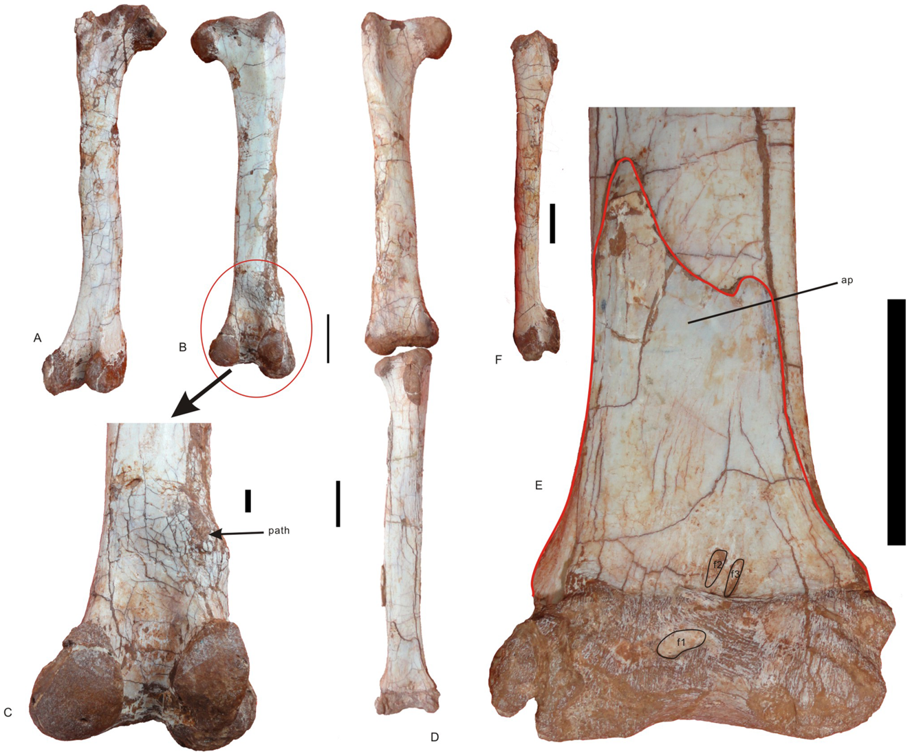
| Описание | Femur, tibia, and proximal tarsals of Nankangia (GMNH F10003). A, Left femur in posterior view; B, Right femur in posterior view; C, Close up of the distal portion of the right femur in posterior view, showing the pathological area; D, Right femur and tibia in anterior view; E, Close-up of the distal portion of the tibia, the astragalus, and the calcaneum in anterior view; F, Left femur in lateral view. Abbreviations: ap., ascending process of astragalus; cal., calcaneum; f1–f3, fossae 1–3; path., pathological areas. Scale bar = 1 cm in C; 5 cm in other images. |
|---|---|
| Источник | http://www.plosone.org/article/info%3Adoi%2F10.1371%2Fjournal.pone.0080557;jsessionid=8FC963102FC951F02382E42B2E5DA827 |
| Время создания | 2013 |
| Автор или правообладатель | Junchang Lü, Laiping Yi, Hui Zhong, Xuefang Wei — Лицензия: CC BY 2.5 (Creative Commons Attribution 2.5) https://creativecommons.org/licenses/by/2.5 |
| Другие версии файла | — |
Источник файла — сайт Wikimedia Commons, куда он был загружен под одной из свободных лицензий ( https://commons.wikimedia.org/wiki/File:Nankangia_leg_bones.png ). Авторов, работавших над этим файлом см. в истории файла: https://commons.wikimedia.org/w/index.php?title=File:Nankangia_leg_bones.png&action=history


В общем случае в статьях энциклопедии Руниверсалис файлы используются в соответствии со статьёй 1274 Гражданского кодекса Российской Федерации.
История файла
Нажмите на дату/время, чтобы увидеть версию файла от того времени.
| Дата/время | Миниатюра | Размеры | Участник | Примечание | |
|---|---|---|---|---|---|
| текущий | 09:51, 21 ноября 2023 | | 1323 × 1098 (1,22 МБ) | I, Robot (обсуждение | вклад) | == Описание == {{Изображение | описание = <p>Femur, tibia, and proximal tarsals of Nankangia (GMNH F10003). </p> A, Left femur in posterior view; B, Right femur in posterior view; C, Close up of the distal portion of the right femur in posterior view, showing the pathological area; D, Right femur and tibia in anterior view; E, Close-up of the distal portion of the tibia, the astragalus, and the calcaneum in anterior view; F, Left femur in lateral view. Abbreviations: ap., ascending process o... |
Вы не можете перезаписать этот файл.
Использование файла
Следующая страница использует этот файл:
