


Файл:Ultrastructure of choroid epithelium.jpg

Исходный файл (1200 × 1000 пкс, размер файла: 414 КБ, MIME-тип: image/jpeg)
Описание
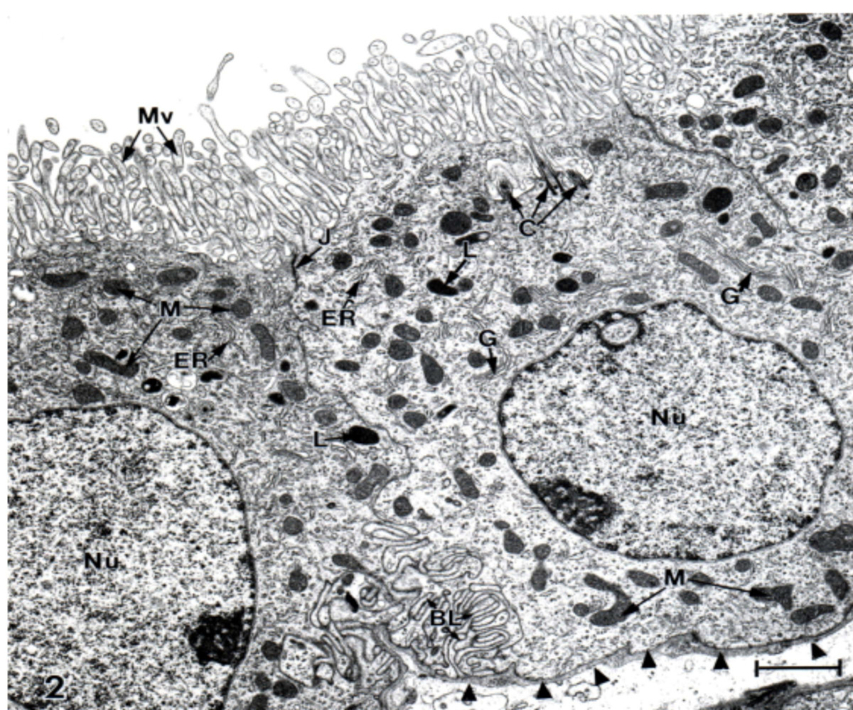
| Описание | Ultrastructure of choroid epithelium. CP from a lateral ventricle of an untreated adult Sprague-Dawley rat was fixed for electron microscopy with OsO4. There is a profusion of apical membrane (CSF-facing) microvilli (Mv) and many intracellular mitochondria (M). J refers to the tight junction welding two cells at their apical poles. C = centriole. G and ER, Golgi apparatus and endoplasmic reticulum. Nucleus (Nu) is oval and has a nucleolus. Arrowheads point to basal lamina at the plasma face of the epithelial cell; the basal lamina separates the choroid cell above from the interstitial fluid below. Basal labyrinth (BL) is the intertwining of basolateral membranes of adjacent cells. Choroidal morphology resembles proximal tubule, consistent with both cell types rapidly turning over fluid. Scale bar = 2 μm. Johanson et al. Cerebrospinal Fluid Research 2008 5:10 doi:10.1186/1743-8454-5-10 |
|---|---|
| Источник | Multiplicity of cerebrospinal fluid functions: New challenges in health and disease |
| Время создания | 2008 |
| Автор или правообладатель | Conrad E Johanson1 , John A Duncan III1 , Petra M Klinge2 , Thomas Brinker2 , Edward G Stopa1 and Gerald D Silverberg — Лицензия: CC BY 2.0 (Creative Commons Attribution 2.0) https://creativecommons.org/licenses/by/2.0 |
| Другие версии файла | — |
Источник файла — сайт Wikimedia Commons, куда он был загружен под одной из свободных лицензий ( https://commons.wikimedia.org/wiki/File:Ultrastructure_of_choroid_epithelium.jpg ). Авторов, работавших над этим файлом см. в истории файла: https://commons.wikimedia.org/w/index.php?title=File:Ultrastructure_of_choroid_epithelium.jpg&action=history


В общем случае в статьях энциклопедии Руниверсалис файлы используются в соответствии со статьёй 1274 Гражданского кодекса Российской Федерации.
История файла
Нажмите на дату/время, чтобы увидеть версию файла от того времени.
| Дата/время | Миниатюра | Размеры | Участник | Примечание | |
|---|---|---|---|---|---|
| текущий | 11:39, 26 января 2024 | | 1200 × 1000 (414 КБ) | Я, робот (обсуждение | вклад) | == Описание == {{Изображение | описание = Ultrastructure of choroid epithelium. CP from a lateral ventricle of an untreated adult Sprague-Dawley rat was fixed for electron microscopy with OsO4. There is a profusion of apical membrane (CSF-facing) microvilli (Mv) and many intracellular mitochondria (M). J refers to the tight junction welding two cells at their apical poles. C = centriole. G and ER, Golgi apparatus and endoplasmic reticulum. Nucleus (Nu) is oval and has a nucleolus. Arrowheads... |
Вы не можете перезаписать этот файл.
Использование файла
Следующий файл является дубликатом этого файла (подробности):

- Файл:Ultrastructure of choroid epithelium.jpg на общем хранилище
Следующая страница использует этот файл:
