


Файл:Nibib 030207 105309 sarcoma.jpg
Nibib_030207_105309_sarcoma.jpg (530 × 400 пкс, размер файла: 88 КБ, MIME-тип: image/jpeg)
Описание
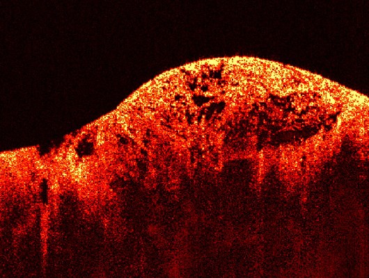
| Описание | Optical Coherence Tomography (OCT) image of a sarcoma
This image of a sarcoma, or muscle tumor, was obtained using Optical Coherence Tomography (OCT). This technique is similar to ultrasound, and yields images of tissues below the surface. The images are on a micron scale, and they are gathered by measuring scattered light waves that bounce off of tissue. In this picture, the tissue looks healthy and normal on the left. To the right, the structure appears cancerous and irregular. Images like this one, obtained by using OCT in real-time, can help to promote human health by detecting tumors early during image-guided procedures, and allowing time for treatment. |
|---|---|
| Источник | NIBIB, http://www.nibib.nih.gov/publicPage.cfm?section=gallery&action=desc&page=1&photo=27 |
| Время создания | Unknown date |
| Автор или правообладатель | Image courtesy of Dr. Stephen Boppart, Biophotonics Imaging Laboratory, University of Illinois at Urbana-Champaign. EB 005221 — Лицензия: Public domain (в общественном достоянии) |
| Другие версии файла | — |
Источник файла — сайт Wikimedia Commons, куда он был загружен под одной из свободных лицензий ( https://commons.wikimedia.org/wiki/File:Nibib_030207_105309_sarcoma.jpg ). Авторов, работавших над этим файлом см. в истории файла: https://commons.wikimedia.org/w/index.php?title=File:Nibib_030207_105309_sarcoma.jpg&action=history


В общем случае в статьях энциклопедии Руниверсалис файлы используются в соответствии со статьёй 1274 Гражданского кодекса Российской Федерации.
История файла
Нажмите на дату/время, чтобы увидеть версию файла от того времени.
| Дата/время | Миниатюра | Размеры | Участник | Примечание | |
|---|---|---|---|---|---|
| текущий | 18:29, 22 ноября 2023 | | 530 × 400 (88 КБ) | I, Robot (обсуждение | вклад) | == Описание == {{Изображение | описание = Optical Coherence Tomography (OCT) image of a sarcoma This image of a sarcoma, or muscle tumor, was obtained using Optical Coherence Tomography (OCT). This technique is similar to ultrasound, and yields images of tissues below the surface. The images are on a micron scale, and they are gathered by measuring scattered light waves that bounce off of tissue. In this picture, the tissue looks healthy and normal on the left. To the right, the structure ap... |
Вы не можете перезаписать этот файл.
Использование файла
Следующий файл является дубликатом этого файла (подробности):

- Файл:Nibib 030207 105309 sarcoma.jpg на общем хранилище
Следующая страница использует этот файл:
