


Файл:Human brainstem anterior view 2 description.JPG
Human_brainstem_anterior_view_2_description.JPG (346 × 487 пкс, размер файла: 35 КБ, MIME-тип: image/jpeg)
Описание
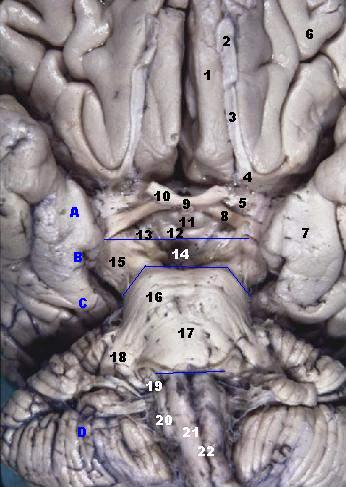
| Описание | Human brainstem anterior view
A: Thalamus, B: Mesencephalon, C: Pons, D: Medulla oblongata
The Medulla, Pons & Midbrain are delineated here on the anterior surface of the brain. In the Midbrain the massive right and left Cerebral Peduncles are seen. The two peduncles are separated by the Interpeduncular Fossa. Posteromedial central arteries enter the brain through the holes in the Posterior Perforated Substance located in the floor of the fossa. Cranial nerve III also enters the midbrain through this fossa. Another important fact to note is that the pituitary gland was removed before this photo was taken. The infundibulum connects between the thalamus and pituitary. (font: arial black, size: 10) |
|---|---|
| Источник | [www.healcentral.org/healapp/showMetadata?metadataId=40566 www.healcentral.org/healapp/showMetadata?metadataId=40566] (Internet Archive of file description page) |
| Время создания | 2005-11-30 |
| Автор или правообладатель | John A Beal, PhD Dep't. of Cellular Biology & Anatomy, Louisiana State University Health Sciences Center Shreveport — Лицензия: CC BY 2.5 (Creative Commons Attribution 2.5) https://creativecommons.org/licenses/by/2.5 |
| Другие версии файла | — |
Источник файла — сайт Wikimedia Commons, куда он был загружен под одной из свободных лицензий ( https://commons.wikimedia.org/wiki/File:Human_brainstem_anterior_view_2_description.JPG ). Авторов, работавших над этим файлом см. в истории файла: https://commons.wikimedia.org/w/index.php?title=File:Human_brainstem_anterior_view_2_description.JPG&action=history


В общем случае в статьях энциклопедии Руниверсалис файлы используются в соответствии со статьёй 1274 Гражданского кодекса Российской Федерации.
История файла
Нажмите на дату/время, чтобы увидеть версию файла от того времени.
| Дата/время | Миниатюра | Размеры | Участник | Примечание | |
|---|---|---|---|---|---|
| текущий | 12:01, 10 августа 2023 | | 346 × 487 (35 КБ) | I, Robot (обсуждение | вклад) | == Описание == {{Изображение | описание = <p><b>Human brainstem anterior view</b> </p> <ol><li>Gyrus rectus</li> <li>Bulbus olfactorii</li> <li>Tractus olfactorii</li> <li>Trigonum olfactorium</li> <li>Substantia perforata anterior</li> <li>Gyri orbitales</li> <li>Gyrus parahippocampalis</li> <li>Tractus opticus</li> <li>Chiasma opticum</li> <li>Nervus opticus</li> <li>Infundibulum hypophysialis</li> <li>Corpora mammillaria</li> <li>Substantia perforata posterior</li> <li>Fossa interpeduncul... |
Вы не можете перезаписать этот файл.
Использование файла
Следующий файл является дубликатом этого файла (подробности):

- Файл:Human brainstem anterior view 2 description.JPG на общем хранилище
Следующие 3 страницы используют этот файл:
