


Файл:Hemangioma of Myometrium (3271483135).jpg

Исходный файл (1456 × 1429 пкс, размер файла: 847 КБ, MIME-тип: image/jpeg)
Описание
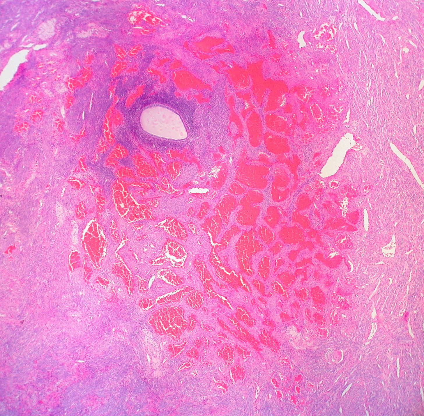
| Описание | This otherwise normal-looking uterus had a 0.3-cm dark purple spot on the cut surface of the myometrium. The island of endometrium seen in association with the hemangioma was solitary. There was no other evidence of adenomyosis. I don't recall ever having seen a hemangioma of the myometrium before. As common as hemangiomas are in other organs, they are rare in the uterus. Pathological and histological images courtesy of Ed Uthman at flickr. |
|---|---|
| Источник |
|
| Время создания | 2009-01-30 13:08 |
| Автор или правообладатель | Ed Uthman from Houston, TX, USA — Лицензия: CC BY 2.0 (Creative Commons Attribution 2.0) https://creativecommons.org/licenses/by/2.0 |
| Другие версии файла | — |
Источник файла — сайт Wikimedia Commons, куда он был загружен под одной из свободных лицензий ( https://commons.wikimedia.org/wiki/File:Hemangioma_of_Myometrium_(3271483135).jpg ). Авторов, работавших над этим файлом см. в истории файла: https://commons.wikimedia.org/w/index.php?title=File:Hemangioma_of_Myometrium_(3271483135).jpg&action=history


В общем случае в статьях энциклопедии Руниверсалис файлы используются в соответствии со статьёй 1274 Гражданского кодекса Российской Федерации.
История файла
Нажмите на дату/время, чтобы увидеть версию файла от того времени.
| Дата/время | Миниатюра | Размеры | Участник | Примечание | |
|---|---|---|---|---|---|
| текущий | 12:17, 22 октября 2023 | | 1456 × 1429 (847 КБ) | Я, робот (обсуждение | вклад) | == Описание == {{Изображение | описание = <p>This otherwise normal-looking uterus had a 0.3-cm dark purple spot on the cut surface of the myometrium. The island of endometrium seen in association with the hemangioma was solitary. There was no other evidence of adenomyosis. </p><p>I don't recall ever having seen a hemangioma of the myometrium before. As common as hemangiomas are in other organs, they are rare in the uterus.<br/></p> Pathological and histological images courtesy of Ed Uthman a... |
Вы не можете перезаписать этот файл.
Использование файла
Следующий файл является дубликатом этого файла (подробности):

- Файл:Hemangioma of Myometrium (3271483135).jpg на общем хранилище
Следующая страница использует этот файл:
