


Файл:Giardia muris trophozoite SEM 11643.jpg

Исходный файл (1496 × 1039 пкс, размер файла: 843 КБ, MIME-тип: image/jpeg)
Описание
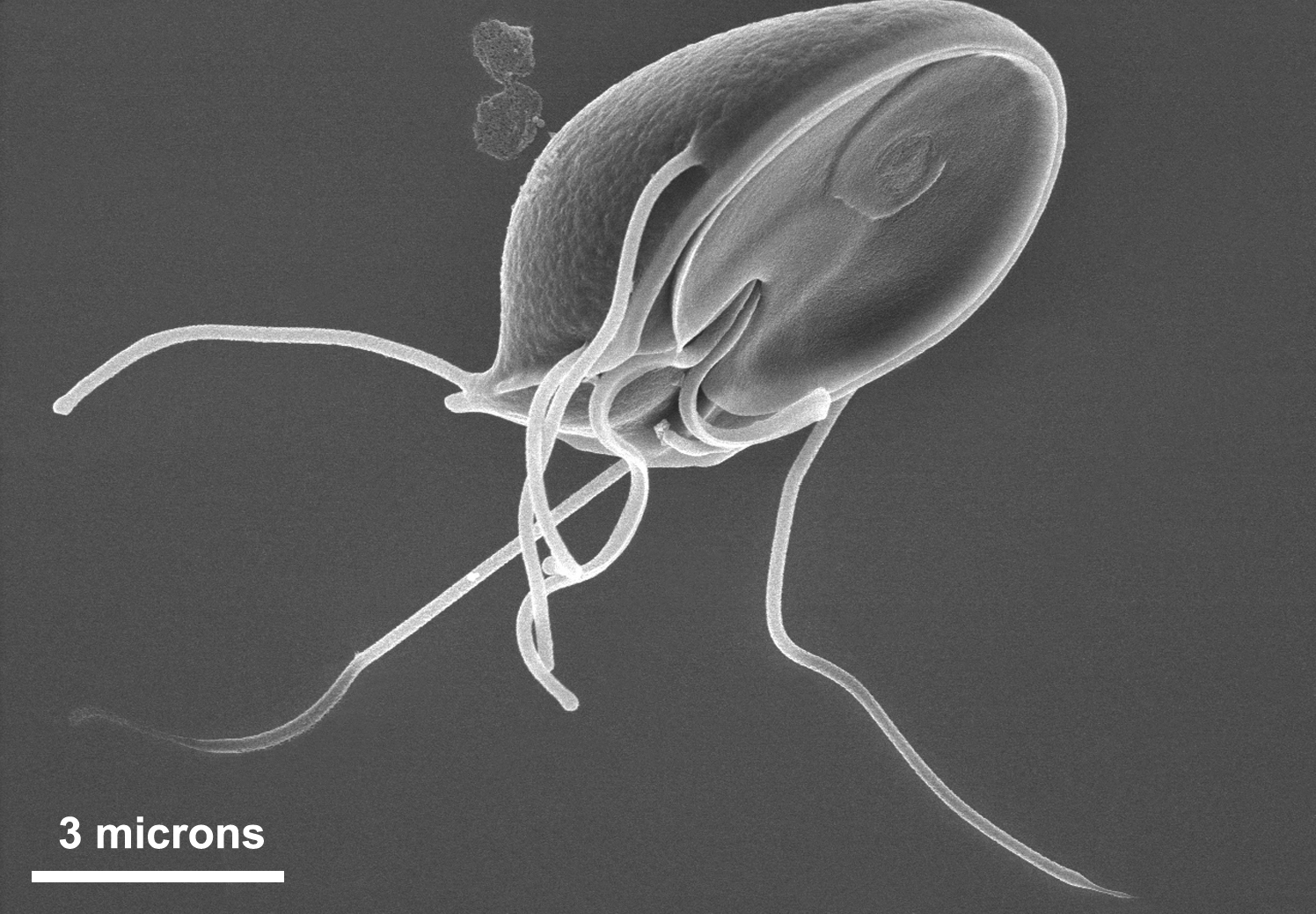
| Описание | This scanning electron micrograph (SEM) clearly showed the ventral surface of a Giardia muris trophozoite. The ventral adhesive disk resembles a suction cup, where overlapping microtubules in the cytoplasm form a number-6-shaped figure. Giardia muris has four pairs of flagella that are responsible for the organism’s motility. The adhesive disk facilitates adherence of the protozoan to the intestinal surface.
The protozoan Giardia causes the diarrheal disease called giardiasis. Giardia species exist as free-swimming (by means of flagella) trophozoites, and as egg-shaped cysts. It is the cystic stage, which facilitates the survival of these organisms under harsh environmental conditions. The cyst is considered the infective form, and disease is often transmitted by drinking contaminated water. As depicted in these SEMs, in the intestine, cysts are stimulated to liberate trophozoites. Cysts can be shed in fecal material, and can, thereafter, remain viable for several months in appropriate environmental conditions. Cysts can also be transferred directly from person-to-person, as a result of poor hygiene. | ||||||||
|---|---|---|---|---|---|---|---|---|---|
| Источник | <tbody>
Источник файла — сайт Wikimedia Commons, куда он был загружен под одной из свободных лицензий ( https://commons.wikimedia.org/wiki/File:Giardia_muris_trophozoite_SEM_11643.jpg ). Авторов, работавших над этим файлом см. в истории файла: https://commons.wikimedia.org/w/index.php?title=File:Giardia_muris_trophozoite_SEM_11643.jpg&action=history В общем случае в статьях энциклопедии Руниверсалис файлы используются в соответствии со статьёй 1274 Гражданского кодекса Российской Федерации. |


История файла
Нажмите на дату/время, чтобы увидеть версию файла от того времени.
| Дата/время | Миниатюра | Размеры | Участник | Примечание | |
|---|---|---|---|---|---|
| текущий | 05:17, 16 октября 2023 | | 1496 × 1039 (843 КБ) | Я, робот (обсуждение | вклад) | == Описание == {{Изображение | описание = This scanning electron micrograph (SEM) clearly showed the ventral surface of a Giardia muris trophozoite. The ventral adhesive disk resembles a suction cup, where overlapping microtubules in the cytoplasm form a number-6-shaped figure. Giardia muris has four pairs of flagella that are responsible for the organism’s motility. The adhesive disk facilitates adherence of the protozoan to the intestinal surface. The protozoan Giardia causes the diarrheal... |
Вы не можете перезаписать этот файл.
Использование файла
Следующий файл является дубликатом этого файла (подробности):

- Файл:Giardia muris trophozoite SEM 11643.jpg на общем хранилище
Следующая страница использует этот файл:
