


Файл:Cytoskeletal organization of dendritic spines (ru).jpg

Исходный файл (1176 × 1280 пкс, размер файла: 418 КБ, MIME-тип: image/jpeg)
Описание
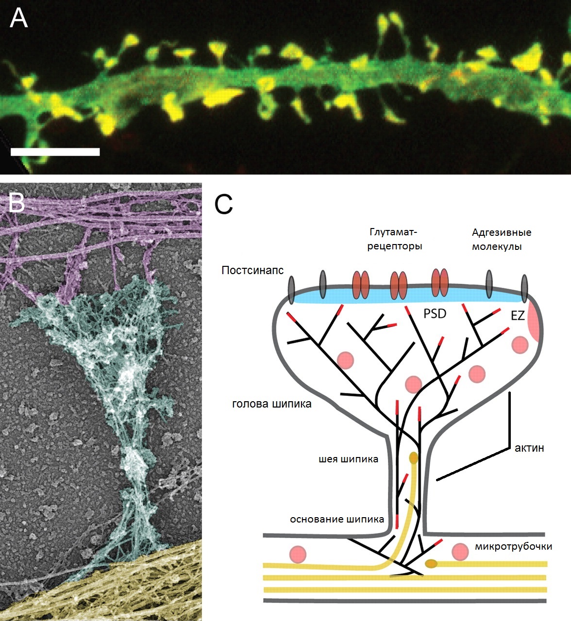
| Описание | Cytoskeletal organization of dendritic spines. (A) Dendritic spine morphology (green) and localization of F-actin (red) in cultured hippocampal neurons. Merged red and green are shown in yellow. Bar, 5 µm. (B) Actin and microtubule cytoskeleton organization in a mature dendritic spine from cultured hippocampal neurons visualized by platinum replica electron microscopy (EM). Axonal cytoskeleton, purple; dendritic shaft, yellow; dendritic spine, cyan. The spine head typically contains a dense network of short cross-linked branched actin filaments, whereas the spine neck contains loosely arranged longitudinal actin filaments, both branched and linear. The base of the spine also contains branched filaments, which frequently reside directly on the microtubule network in the dendritic shaft. (C) Schematic diagram of a mature mushroom-shaped spine showing the postsynaptic membrane containing the postsynaptic density (PSD; blue), adhesion molecules (gray) and glutamate receptors (reddish brown), the actin (black lines) and microtubule (yellow) cytoskeleton, and organelles. The endocytic zone (EZ) is located lateral of the PSD in extrasynaptic regions of the spine and recycling endosomes (pink) are found in the shaft and spines. Dendritic spines exhibit a continuous network of both straight and branched actin filaments (black lines). The actin network is spread in the spine base, gets constricted in the neck, undergoes extensive branching at the neck–head junction, and stays highly branched in the spine head. The actin-polymerizing barbed ends are indicated as red lines. Stable microtubule arrays are predominantly present in the dendritic shaft. A small fraction of the microtubules in mature dendrites are dynamic and depart from the dendritic shaft, curve, and transiently enter dendritic spines. The microtubule plus-ends are symbolized as yellow ovals. |
|---|---|
| Источник | File:Cytoskeletal_organization_of_dendritic_spines.jpg |
| Время создания | 2013-10-22 |
| Автор или правообладатель | Hotulainen P, Hoogenraad CC — Лицензия: CC BY-SA 2.5 (Creative Commons Attribution-Share Alike 2.5) https://creativecommons.org/licenses/by-sa/2.5 |
| Другие версии файла | — |

Источник файла — сайт Wikimedia Commons, куда он был загружен под одной из свободных лицензий ( https://commons.wikimedia.org/wiki/File:Cytoskeletal_organization_of_dendritic_spines_(ru).jpg ). Авторов, работавших над этим файлом см. в истории файла: https://commons.wikimedia.org/w/index.php?title=File:Cytoskeletal_organization_of_dendritic_spines_(ru).jpg&action=history
.jpg)
.jpg&action=history)
В общем случае в статьях энциклопедии Руниверсалис файлы используются в соответствии со статьёй 1274 Гражданского кодекса Российской Федерации.
История файла
Нажмите на дату/время, чтобы увидеть версию файла от того времени.
| Дата/время | Миниатюра | Размеры | Участник | Примечание | |
|---|---|---|---|---|---|
| текущий | 20:17, 4 октября 2023 | | 1176 × 1280 (418 КБ) | Я, робот (обсуждение | вклад) | == Описание == {{Изображение | описание = Cytoskeletal organization of dendritic spines. (A) Dendritic spine morphology (green) and localization of F-actin (red) in cultured hippocampal neurons. Merged red and green are shown in yellow. Bar, 5 µm. (B) Actin and microtubule cytoskeleton organization in a mature dendritic spine from cultured hippocampal neurons visualized by platinum replica electron microscopy (EM). Axonal cytoskeleton, purple; dendritic shaft, yellow; dendritic spine, cyan.... |
Вы не можете перезаписать этот файл.
Использование файла
Следующий файл является дубликатом этого файла (подробности):
.jpg)
- Файл:Cytoskeletal organization of dendritic spines (ru).jpg на общем хранилище
Следующая страница использует этот файл:
.jpg&oldid=17600237)