


Файл:A computed tomography brain scan showing bilateral basal ganglia calcification.jpg

Исходный файл (1200 × 883 пкс, размер файла: 153 КБ, MIME-тип: image/jpeg)
Описание
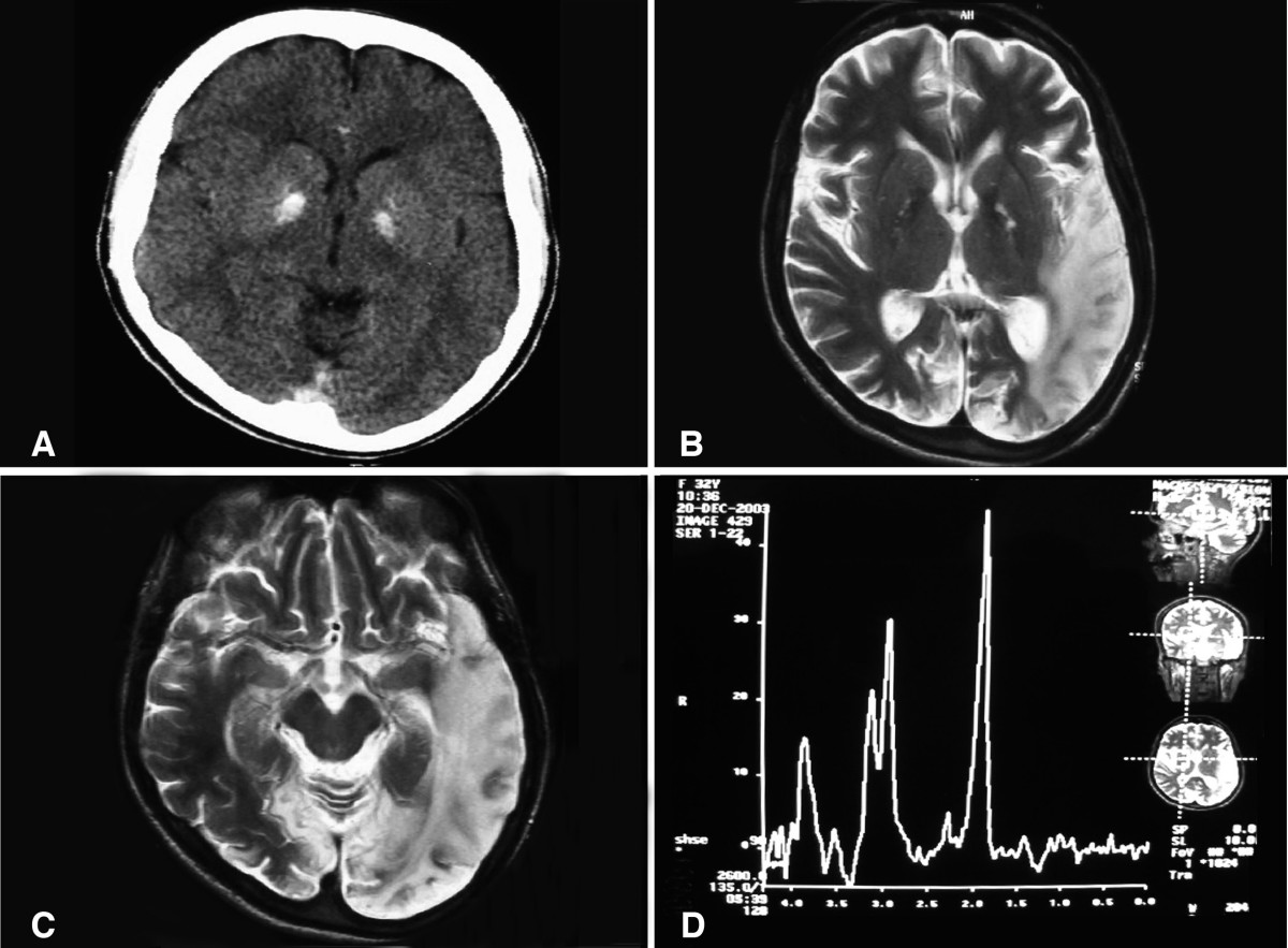
| Описание | (a) A computed tomography brain scan showing bilateral basal ganglia calcification; the cerebellum shows prominent folia indicating mild cerebellar atrophy. (b) Axial T2 brain magnetic resonance image scan showing left temporo-parieto occipital ischemic lesion. (c) Axial T2 brain magnetic resonance image scan showing the extension of the parietal temporal region to the occipital lobe, and also showing a right occipital lesion. (d) Magnetic resonance spectroscopy showing inversion of J-coupling phenomenon at 1.3 ppm, indicating lactate peak. Abu-Amero et al. Journal of Medical Case Reports 2009 3:77 doi:10.1186/1752-1947-3-77 |
|---|---|
| Источник | A patient with typical clinical features of mitochondrial encephalopathy, lactic acidosis and stroke-like episodes (MELAS) but without an obvious genetic cause: a case report |
| Время создания | 2009 |
| Автор или правообладатель | Abu-Amero KK, Al-Dhalaan H, Bohlega S, Hellani A, Taylor RW. — Лицензия: CC BY 2.0 (Creative Commons Attribution 2.0) https://creativecommons.org/licenses/by/2.0 |
| Другие версии файла | — |
Источник файла — сайт Wikimedia Commons, куда он был загружен под одной из свободных лицензий ( https://commons.wikimedia.org/wiki/File:A_computed_tomography_brain_scan_showing_bilateral_basal_ganglia_calcification.jpg ). Авторов, работавших над этим файлом см. в истории файла: https://commons.wikimedia.org/w/index.php?title=File:A_computed_tomography_brain_scan_showing_bilateral_basal_ganglia_calcification.jpg&action=history


В общем случае в статьях энциклопедии Руниверсалис файлы используются в соответствии со статьёй 1274 Гражданского кодекса Российской Федерации.
История файла
Нажмите на дату/время, чтобы увидеть версию файла от того времени.
| Дата/время | Миниатюра | Размеры | Участник | Примечание | |
|---|---|---|---|---|---|
| текущий | 02:52, 6 сентября 2023 | | 1200 × 883 (153 КБ) | I, Robot (обсуждение | вклад) | == Описание == {{Изображение | описание = (a) A computed tomography brain scan showing bilateral basal ganglia calcification; the cerebellum shows prominent folia indicating mild cerebellar atrophy. (b) Axial T2 brain magnetic resonance image scan showing left temporo-parieto occipital ischemic lesion. (c) Axial T2 brain magnetic resonance image scan showing the extension of the parietal temporal region to the occipital lobe, and also showing a right occipital lesion. (d) Magnetic resonance... |
Вы не можете перезаписать этот файл.
Использование файла
Следующий файл является дубликатом этого файла (подробности):

Следующая страница использует этот файл:
